Harvard Study Finds Genetic ‘Toggle Switch’ for Sociability
Harvard Study Finds Genetic ‘Toggle Switch’ for Sociability
Study pinpoints genes and neural circuitry behind autism's impaired sociability.

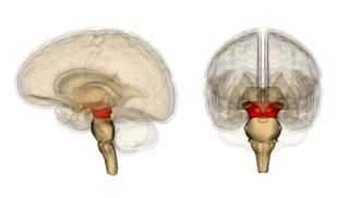
Midbrain (in red) is the seat of the ventral tegmental area (VTA) which may house a genetic 'toggle switch' for sociability.
Source: Wikimedia Commons/Life Sciences Database
Using "chemogenetic" tools in living mice, the researchers found that increasing the UBE3A gene downregulates a glutamate-based synapse organizer called “CBLN1,” which appears to inhibit sociability in mice, and presumably humans. The March 2017 findings were published today in the journal Nature.
The research team, led by Matthew P. Anderson, Associate Professor in the Departments of Pathology and Neurology and Director of Neuropathology at BIDMC, was able to pinpoint how UBE3A influences the neurobiological control of sociability like a ‘toggle switch’ through complex neuronal mechanisms.
In previous research, Anderson's team demonstrated that mice engineered with extra copies of the UBE3A gene displayed impaired sociability. Notably, the lack of the UBE3A gene in humans is linked to a rare genetic and neurological disorder called "Angelman syndrome" (AS), which is characterized by exuberant sociability and other symptoms.
In 1965, Dr. Harry Angelman first identified Angelman syndrome, which produces symptoms such as the inability to coordinate voluntary movements (ataxia), jerky movements of the arms and legs; as well as increased sociability, a joyful disposition, and unprovoked episodes of laughter or smiling. The fine-tuned movement and balance impairments observed in Angelman syndrome are widely considered to be linked to deficits in the cerebellum (Latin for "Little brain").
After analyzing and comparing interactions between the UBE3A genes and other genes altered in human autism, the researchers noticed that increased amounts of UBE3A repressed a family of genes known as “Cerebellin genes.” These genes interact with other autism genes to form glutamatergic synapses in the junctions where neurons communicate via the neurotransmitter glutamate.
Anderson et al. chose to focus specifically on Cerebellin 1 (CBLN1), as the potential mediator of UBE3A's effects. When they deleted CBLN1 in glutamate neurons, they recreated the same impaired sociability produced by increased UBE3A.
Other studies have found that CBLN1 is required for synapse integrity and synaptic plasticity during synapse formation in the cerebellum. CBLN1 is also essential for the formation and maintenance of parallel fibers and Purkinje cell synapses in the cerebellum. In a statement to BIDMC, Anderson said:
"Selecting Cerebellin 1 out of hundreds of other potential targets was something of a leap of faith. When we deleted the gene and were able to reconstitute the social deficits, that was the moment we realized we'd hit the right target. Cerebellin 1 was the gene repressed by UBE3A that seemed to mediate its effects."For their latest study, the BIDMC researchers conducted various brain mapping experiments to pinpoint where the gene interactions linked to sociability were taking place in the brain. Through a variety of experiments, they honed in on the ventral tegmental area (VTA) midbrain section of the brainstem.
Chemogenetic techniques allowed the researchers to switch a specific group of neurons in the VTA on and off. By turning these neurons on, the researchers increased sociability. Conversely, turning these neurons off reduced sociability. Interestingly, the VTA is commonly considered a reward center that plays a role in addiction and substance abuse.
In his statement to BIDMC, Anderson elaborated on the findings of his latest study:
"In this study, we wanted to determine where in the brain this social behavior deficit arises and where and how increases of the UBE3A gene repress it. We had tools in hand that we built ourselves. We not only introduced the gene into specific brain regions of the mouse, but we could also direct it to specific cell types to test which ones played a role in regulating sociability.
We mapped this seat of sociability to a surprising location. Most scientists would have thought they take place in the cortex—the area of the brain where sensory processing and motor commands take place—but, in fact, these interactions take place in the brain stem, in the reward system.
We were able to abolish sociability by inhibiting these neurons and we could magnify and prolong sociability by turning them on. So we have a toggle switch for sociability. It has a therapeutic flavor; someday, we might be able to translate this into a treatment that will help patients."
When Anderson and colleagues compared the brains of the mice engineered to model autism to those of normal ("wild type") mice, they observed that the increased UBE3A gene copies interacted with almost 600 other genes.
In another series of experiments, Anderson and colleagues demonstrated an even more definitive link between UBE3A and CBLN1 as it related to seizures. Anderson and colleagues found that deleting UBE3A (upstream from cerebellin genes) prevented the seizure-induced social impairments and the ability of a seizure to repress CBLN1.
In their study, the authors write, “Seizure-induced deficits are UBE3A-dependent. CBLN1 is also repressed by increased neuronal activity in the cerebellum in culture after depolarization and in vivo after status epilepticus. Therefore, we tested whether recurrent seizures can concurrently repress CBLN1 and sociability.”
Seizures are a common symptom among people with the UBE3A-related type of autism. When seizures are severe, they also impair sociability. Anderson's team suspected this seizure-induced impairment of sociability was the result of repressing the Cerebellin genes. The researchers conclude that genetic interactions in VTA glutamatergic neurons may impair sociability by downregulating CBLN1.
"If you take away UBE3A, seizures can't repress sociability or Cerebellin," Anderson said. "The flip side is, if you have just a little extra UBE3A—as a subset of people with autism do—and you combine that with less severe seizures, you can get a full-blown loss of social interactions."
The groundbreaking new findings by Anderson and his colleagues at BIDMC advance our understanding of the neuronal circuitry that drives sociability. Hopefully, future research on UBE3A and CBLN1 will lead to targeted interventions that can help those with autism spectrum disorders and Angelman syndrome improve atypical sociability-related symptoms.

